Blood Vessels
Pathway for blood which is the medium of transport that carries nutrients, gases and wastes.
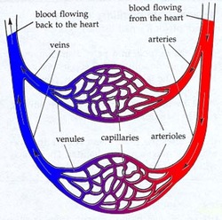
Arteries- Oxygenated blood away from heart, thick walls to withstand blood pressure, elastic muscle fibres, highest blood pressure, smooth muscle to regulate size. (10% of blood)
Arterioles- Connect arteries to capillaries, smaller than arteries, can dilate or constrict to regulate blood pressure.
Veins- De-oxygenated blood back to heart, thinner walls, contains valves to prevent back flow, lowest blood pressure (70% of blood)
Venules- Connect veins to capillares, very thin walls.
Capillaries- Narrow, microscopic tubes, blood cell pass in single file, site of nutrient/waste exchange, thin walls to allow for diffusion, large surface area for greater exchange of gases and nutrients, largest cross-sectional area.
Blood Plasma- Makes up 55% of blood, 90% of plasma is water with dissolved substance. Contains: water, blood proteins, albumin(osmotic blance) + fibrinogen (blood clotting), gases, nutrients, wastes, vitamins, hormones, salts.
1. Red Blood Cells- most plentiful, biconcave disc, no nucleus, live for 120 days, in cell is hemoglobin; binds to oxygen.
2. White Blood Cells- largest, contain nuclei, fight infection, some phagocytic
3. Platelets, very small irregular cell fragment shape, blood clotting.
(All made in bone marrow)
Antigen- (bad) an agent that is foreign or self and is recognized by the immune system.
Antibody- (good) a Y shaped protein capable of identifying and binding to a specific antigen.
Pulmonary-From heart to lungs then back to heart.
-Oxygenated blood to heart. -De-oxygenated blood (CO2) out. |
Systemic-From heart to every cell of body and back to heart.
-For cellular respiration. -De-oxygenated blood back to heart (O2 diffuses out of blood CO2 diffuse in. |
Fetal 4
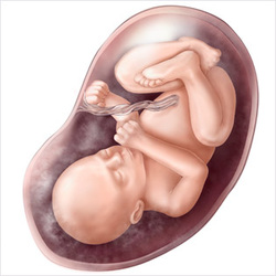
No pulmonary circuit, the placenta is the organ responsible for delivery of nutrients, oxygenated blood and removal of wastes through diffusion and active transport.
1. Umbilical Cord- 1 vein for oxygenated/nutrient rich blood. 2 arteries for de-oxygenated blood from iliac arteries out to plancenta.
2. Venous Duct- Liver bypass moves blood into fetal system.
2. Foramen Ovale- Opening between left and right atrium to bypass lungs.
4. Ductus Arteriosus- Vessel that connects pulmonary artery to aorta.
Capillary Fluid Exchange
- Arteriole side blood pressure higher than osmotic pressure.
- High pressure pushes oxygen/nutrients into surrounding tissue.
- Blood proteins and blood cells are too big to stay in capillaries.
- Oxygen, glucose, amino acid diffuse into cells.
- CO2 and ammonia produced in tissue diffuse out
- Blood pressure reduced less than osmotic pressure.
- Fluid containing waste moved into blood vessels
- Remaining fluid enters lymph vessels carried back to circulatory system
Lymph System
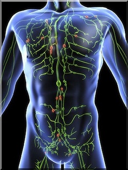
-One way transport system.
-Takes up excess tissue fluid and returns it to the blood.
-Fluid called lymph.
-Transport fatty acids.
-Fight infections (lymphocytes)
-Join blood at subclavian
Parts:
1. Lacteal- dead end of lymph vessels in villi of the small intestine
2. Lymph nodes- filter lymph, waste, dead cells (oval structures)
3. Spleen- stores blood
4. Thymus gland- production and matuation of some lymphocytes
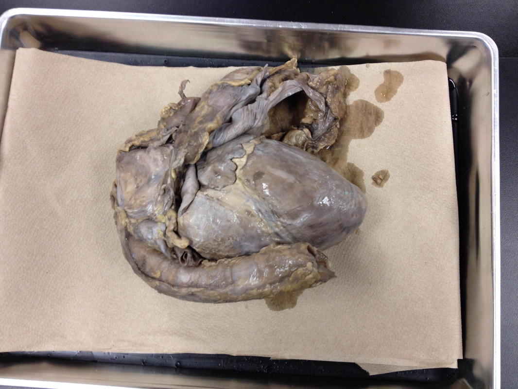
Heart Structure

(P) SA Node- upper wall, pace maker, activates atria
(QRS) AV Node- connect of bundle of HIS, contraction of both ventricles via purkinje fibers
SA node is connected to the brain via the Vagus nerve. The medulla oblongata part of brain that monitors blood pressure. Regulation is autonomic.
GRAPH ---->
ekg- measures electrical activity of heart electrical activity of heart in millivolts (mV). The heart beat is 50-80 beats/min
Systolic Pressure- pressure created when ventricles contract and blood forced against blood vessel.
Diastolic Pressure- pressure created from atrial contraction, measured along brachial artery.
Hypertension- High blood pressure
Hypotension- Low blood pressure

|
|
